Anatomy of the proximal anterior upper leg as illustrated in Pernkopf's
4.9 (528) In stock

Download scientific diagram | Anatomy of the proximal anterior upper leg as illustrated in Pernkopf's atlas. Figure a: Upper part of the illustration selected by (Yee, Coombs, et al. 2018) (first published in 1941 as figure 188 in volume II of Pernkopf's original German edition). The inlay shows the complete illustration (with an SS symbol in the signature). The fascia lata and the fasciae of the muscles are removed. Two nerves of equal diameter (arrowheads) exit the pelvis and join anterior to the sartorius muscle to form the lateral femoral cutaneous nerve (lf). This situation is not to be expected in the average individual -it is a very rare variation. Note how the lateral femoral cutaneous nerve continues as an essentially single nerve stem that gives rise to several small branches. Figure b in the atlas precedes a. In Figure b, the fasciae covering the muscles are still intact except for the slit anterior fascial sheet of the sartorius muscle. Note the missing stem of the lateral femoral cutaneous nerve, which according to a and the branches plotted there would be expected in the marked area. (Since in this case the original paintings are missing, the scans shown here were made using the Josephinum's collection of proofs or test prints as a substitute; image credit: Medical University of Vienna, MUW-AD-3250-5-336-11-Seite-1 and MUW-AD-3250-5-337-1. from publication: What Should Be Done with Pernkopf’s Anatomical Illustrations? A Commentary from the Medical University of Vienna | Thanks to a recent donation by Elsevier, the Medical University of Vienna now holds in its collections the known existing original paintings for Eduard Pernkopf's Atlas of Topographic and Applied Human Anatomy. This atlas is widely considered a pinnacle of the art of | Illustration, Medicalization and Atlas | ResearchGate, the professional network for scientists.
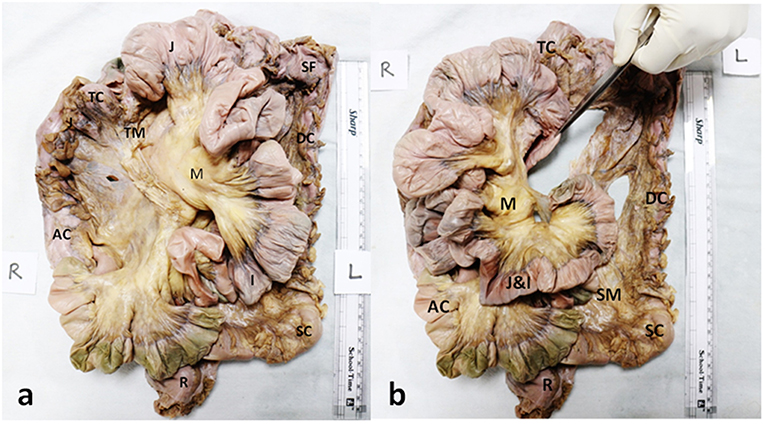
Frontiers Development of a Novel Technique to Dissect the Mesentery That Preserves Mesenteric Continuity and Enables Characterization of the ex vivo Mesentery

The bones of the upper leg and kneecap (patella). (a) The femur
The Skeletomotor System (Chapter 12) - Handbook of Psychophysiology

Persistence of the Ischiadic Artery
Pernkopf Anatomy: Head and neck - Eduard Pernkopf - Google Books

PDF) The blood supply to the sacrotuberous ligament

Pernkopf's Atlas of Topographical and Applied Human Anatomy A detailed and precise anatomical atlas
Codex 99

View of The History of Eduard Pernkopf's Topographische Anatomie des Menschen
Muscles of the upper leg, illustration Stock Photo - Alamy
BIO 112 - Muscles of the Upper Leg Quiz - By tgardiner9
Thigh pain Treatment: Upper, Outer & Inner thigh muscle injuries
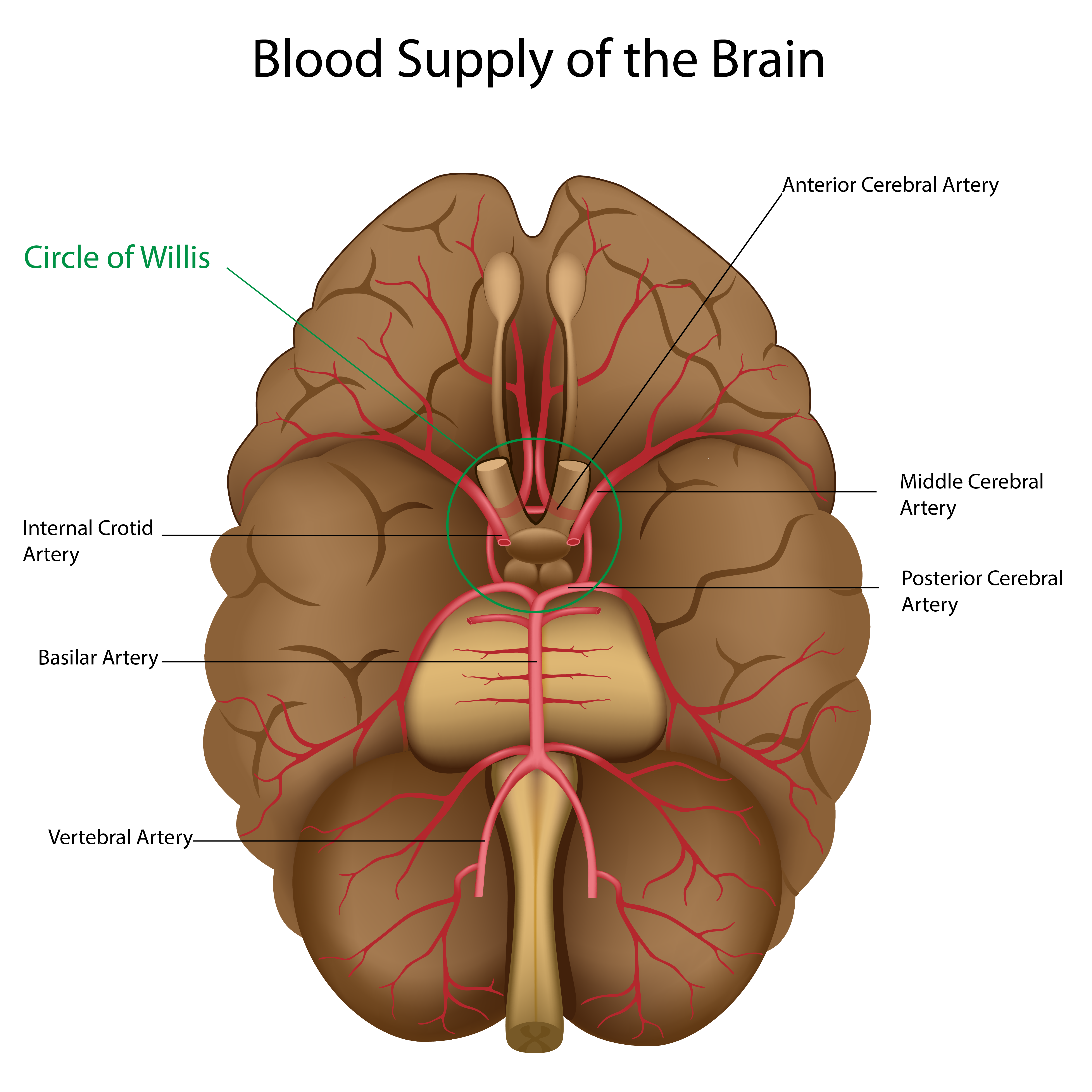 Blood Flow Through the Circle of Willis
 Classy Style. Man Bearded Hipster Wear Classic Suit Outfit. Formal Outfit. Take Good Care of Suit Stock Image - Image of formal, director: 158888887
 Free People Solid Rib Brami
 Genérico Pantalones Termicos Mujer Invierno Pantalones Baggy Mujer PantalóN Jogger Mujer Pantalon Trekking Mujer Pantalones Yoga Mujer PantalóN De
 Oasis Bath Towel in Atlantic Blue
 Women's Board Shorts - Seafolly & Sunseeker Board Shorts